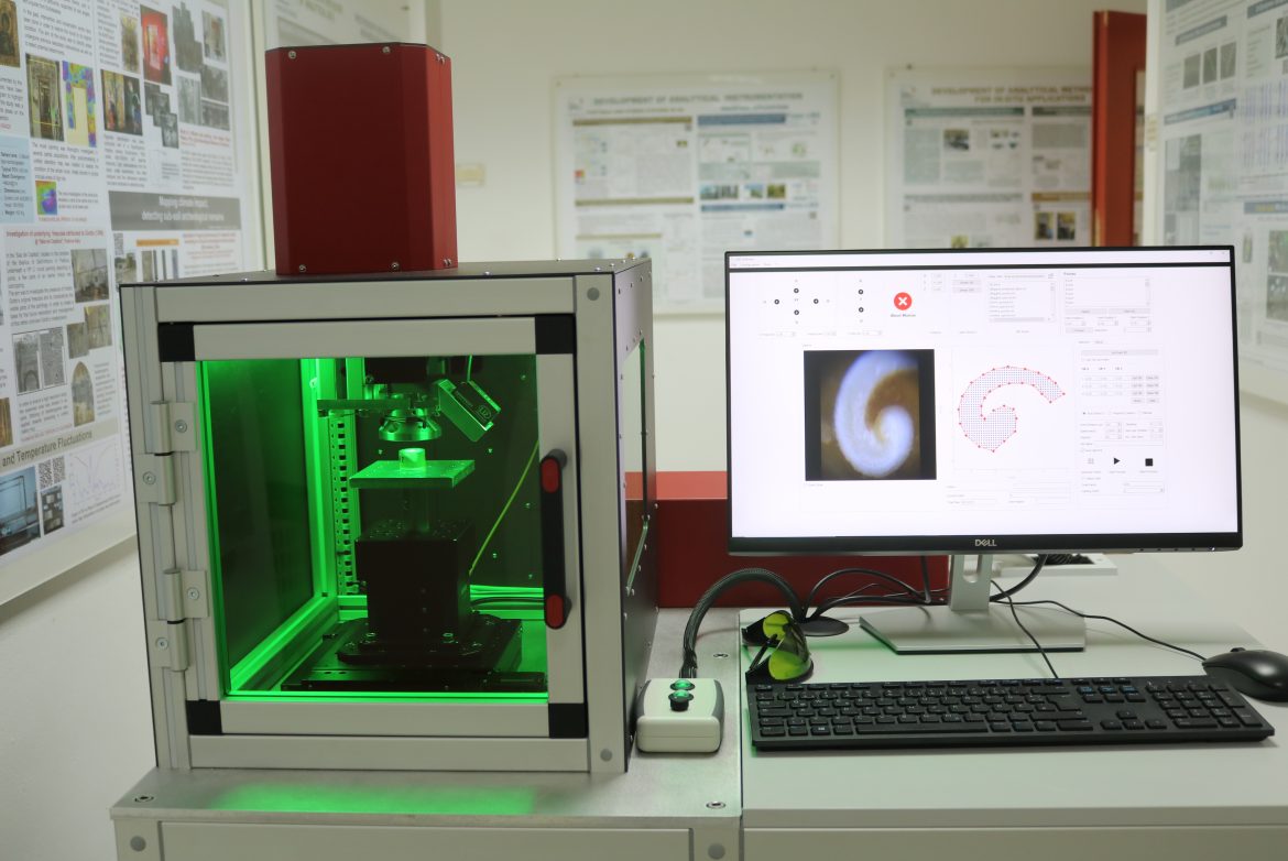
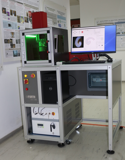
Micro-LIBS workstation, Laser spectroscopy
The LIBS microscope is capable of providing with a fast elemental mapping of flat surfaces, typical cross-sections of geological samples, marine shells, bones, teeth, etc. The 2D-elemental maps of the scanned surface can be used to identify the distribution of mineral phases in rocks, measure the variability of elementalproxies related to the paleoenvironment in shells, and to assess the diffusion of environmental pollutants into hard tissues.
More details of LIBS technique can be found here
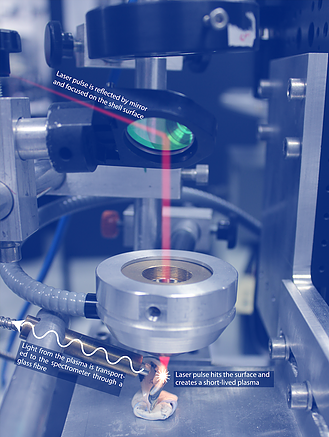
Our prototype micro-LIBS workstation uses a Q-switched nanosecond Nd:YAG laser (λ = 1064 nm, pulse energy 5-20 mJ) in combination with XYZ motorized stages to obtain 2D elemental maps. The laser beam is focused on the sample through a laser objective lens down to a spot of 40 – 60 μm in diameter. A CCD camera allows the user to view the sample surface and define the mapping area. For the LIBS signal acquisition a dual spectrometer unit is employed (AvaSpec-2048-2-USB2, spectral range: 200 – 640 nm, resolution ~ 0.2 – 0.3 nm). If higher sensitivity is needed, the signal can be recorded by a Czerny-Turner spectrometer (Jobin Yvon, TRIAX 320) with an ICCD camera (DH734–18F, Andor Technology) (spectral range ~ 45 nm). With this prototype system a 2D map of 1000 positions, with a distance between points of 100 μm and 10 spectra per analyzed position, can be generated in approximately 30 minutes.
A new, advanced micro-LIBS system was developed in collaboration with Dr. Niklas Hausmann and Danae Theodoraki at the Leibniz-Zentrum für Archäologie, Mainz, Germany (LEIZA). This system uses a diode pumped laser working at a repetition rate of 100 Hz, completely synchronized with the sample stages’ movement that allows the scanning of a whole shell (approx. 5000 points, 10 shots per position) in around 15 minutes.
The entire scanning process (sample movement, auto-focus, laser firing, spectra acquisition, etc.) is controlled by custom-made software specifically developed for this work. This software includes several user-friendly features for accurately setting up the scanning area (sample visualization and definition of the area to scan) and post-processing of the acquired spectra (2D elemental maps generation and visualization).
The LIBS instrument developed and built at IESL-FORTH (Greece) was delivered to the Römisch-Germanisches Zentralmuseum, the German research museum, where it is being employed to analyze thousands of archeological shells.
Publications
N. Hausmann, A.L. Prendergast, A. Lemonis, et al. Extensive elemental mapping unlocks Mg/Ca ratios as climate proxy in seasonal records of Mediterranean limpets. Sci Rep 9, 3698 (2019). DOI: https://doi.org/10.1038/s41598-019-39959-9
N. Hausmann, H.K. Robson and C. Hunt. Annual Growth Patterns and Interspecimen Variability in Mg/Ca Records of Archaeological Ostreaedulis (European Oyster) from the Late Mesolithic Site of Conors Island. Open Quaternary, 5(1), p.9 (2019). DOI: http://doi.org/10.5334/oq.59
N. Hausmann, P. Siozos, A. Lemonis, A. C. Colonese, H. K. Robson and D. Anglos. Elemental mapping of Mg/Ca intensity ratios in marine mollusk shells using laser-induced breakdown spectroscopy. J. Anal. At. Spectrom. 32, p.1467 (2017). DOI: https://doi.org/10.1039/C7JA00131B
Funding
Marie Skłodowska Curie’s actions support the mobility of researchers within and beyond Europe. MSCA is solely funding the ACCELERATE Individual Fellowship.
ACCELERATE is a project funded by the European Union, H2020-MSCA-IF-2015, Marie Curie Intra-European Fellowship, under GA No. 703625. Views and opinions expressed are however those of the authors only and do not necessarily reflect those of the European Union. Neither the European Union can be held responsible for them.
New Emmy Noether Group | SeaFront (seafront-project.com)
The contents of the website reflect only the author’s view and the Agency and the European Commission is not responsible for any use that may be made of the information it contains.