
Spectral Imaging
The technique Spectral Imaging allows the detailed mapping of the individual layers that compose an artwork using a prototype spectral imaging system. The artwork can be thoroughly examined in a wide range of spectral bands (350 nm– 1200 nm) expanding thus the imaging potential of the object to spectral areas in which human eye is not sensitive to; namely the ultraviolet (350 nm- 400 nm) and the near-infrared (700 nm-1200 nm).
In these spectral ranges light penetrates matter in variable ways (less in UV and more in the IR) revealing information for the superficial or for the underlying layers of the painting. In addition, in the visible region spectral imaging enables the analysis with higher spectra sensitivity (13 bands) than the human eye (3 bands), assisting on the differentiation of pigments with similar color perception.
Consequently, the artworks can be studied stratigraphically (differentiating between the individual paint layers), revealing information on their history, composition and structure as well as on their restoration interventions. Information on hidden under-drawings and the series that the pigments were applied allows art historians and scholars to draw conclusions about the period that the artwork was created and the artists’ technique and potentially verify the authenticity of an artwork.
Furthermore, the presence of superficial layers and/or retouches contributes to the knowledge and understanding of their history, previous interventions and their preservation state and thus will assist the conservators to decide on the necessary conservation strategies.
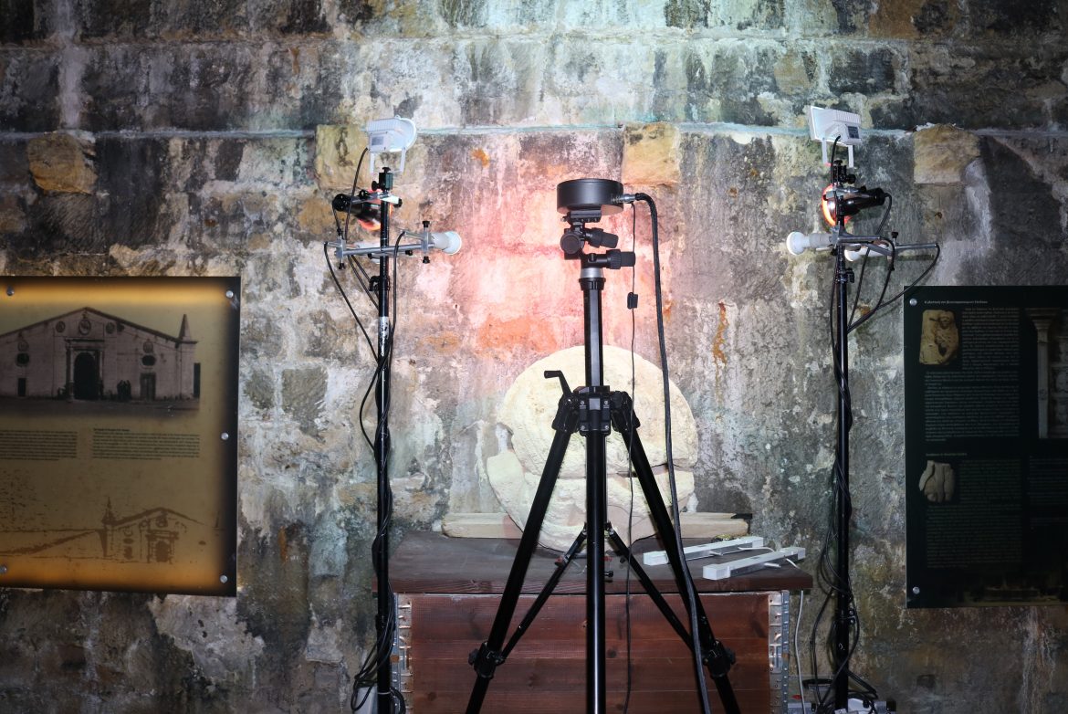
The prototype spectral imaging system IRIS-I It is important to mention that IRIS-I is a portable system, allowing the direct examination of artworks without its transportation into a conservation laboratory.
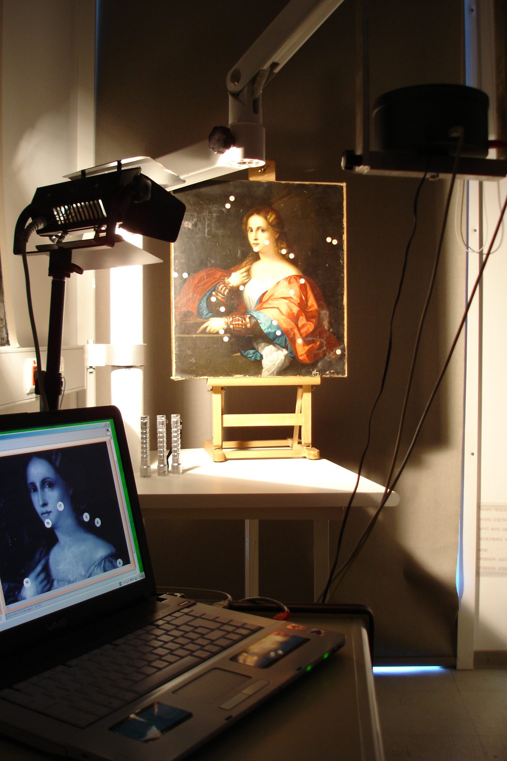 The prototype spectral imaging system IRIS-I It is important to mention that IRIS-I is a portable system, allowing the direct examination of artworks without its transportation into a conservation laboratory.
Imaging in the visible region of light
Imaging artworks in the visible region serves for documentation purposes, reference and for pigment differentiation due to higher spectral resolution
UV induced fluorescence imaging in the visible
This application refers to the recording of the fluorescence originating from the surface layers of the artwork which contain or consist of materials, which fluoresce upon ultraviolet light excitation. This enables the identification and differentiation of protective layers (i.e. natural varnishes and synthetic coatings) and the mapping of biological colonies (i.e. fungi). Finally, it is possible to reveal over-drawings, retouches, or other restoration interventions.
Ultraviolet reflection imaging
Reflection imaging in the ultraviolet (<400nm) provides information regarding the superficial layers of the artwork. Studying artworks in this spectral region reveals the presence of varnishes and other synthetic protective coatings while giving information on their preservation state and informs on superficial deposits. Finally, it is possible to map any retouches or over-paintings referring to later interventions.
Imaging in the near infrared light region
Studying artworks in the infrared region (>700nm) it is possible to get information on the deeper pigment layers. This is possible due to the property of infrared light to penetrate the surface layers of the artwork, allowing light to be absorbed or scattered from deeper layers according to their chemical composition. The recording and imaging of the artwork in this spectral region can provide information related to its deeper paint layers as for example preparatory drawings, modifications of the original composition made by the artist during the process of painting the work (pentimenti), or even to reveal hidden underlying paintings, etc.